Time: 2024-07-26
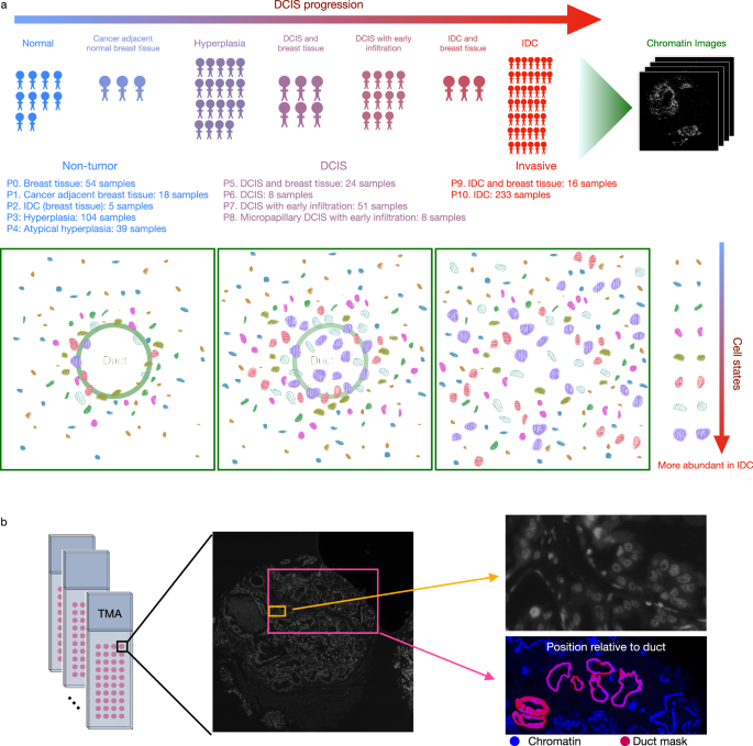
A study conducted on tissue microarrays has shed new light on the analysis of Breast cancer , particularly focusing on Ductal Carcinoma In Situ ( DCIS ) . The research utilized chromatin staining to generate a comprehensive dataset for comparison of different phenotypic categories in non - tumor , DCIS , and invasive ductal carcinoma ( IDC ) patients . The study involved imaging 560 tissue microarray samples from 122 patients at various disease stages and phenotypic categories annotated by pathologists . The tissue microarrays were stained with protein markers in addition to chromatin staining . The study identified distinct cell clusters based on nuclear morphometrics and chromatin organization using a machine - learning framework , revealing differences in cell states across disease stages and phenotypic categories.
The research further explored the pseudo - time ordering of cells in the autoencoder latent space and observed a correlation between the enrichment of cell states and disease stages . The study demonstrated that unsupervised features learned by the autoencoder from chromatin images provided interpretable nuclear and chromatin morphometric features that could explain differences in cell states . Additionally , the position of cells relative to breast ducts was found to be dependent on cell state and disease stage , with differences in proximity to ducts observed between healthy and malignant cell states.
Moreover , the study analyzed the co - localization pattern of cell states and found it to be predictive of disease stage and phenotypic category . Interestingly , the spatial organization of cell states was more informative than cell state abundance for accurately classifying hyperplasia , DCIS , and IDC . The study also highlighted the importance of cell state co - localization patterns in distinguishing between hyperplasia , atypical hyperplasia , DCIS , and breast tissue . Overall , the findings suggest that the spatial organization of cell states defined by chromatin staining could potentially aid in the classification of different phenotypic categories of breast cancer.
In conclusion , the study provides valuable insights into the analysis of breast cancer using high - resolution chromatin imaging datasets . The utilization of Artificial intelligence in analyzing cell states and their spatial organization has the potential to enhance the understanding and classification of different stages of breast cancer , ultimately contributing to improved diagnosis and treatment strategies.


 Business
